
Briefly about Structural Biology
The very core of life lies in the interaction between atoms in molecules. Proteins interacting with each other or with DNA/RNA. Lipids forming membranes holding proteins in place to facilitate transport across cell membranes. To name just a few. To understand the nature of these interactions and how the life of the organism in which they reside changes upon disturbance of these, several techniques are offered within the SciLifeLab capability Integrated Structural Biology. Using only one technique does not always answer the scientific question, and we offer support to have projects integrated between techniques.
The techniques that come together within the capability Integrated Structural Biology can guide the scientist through:
- Interaction of small ligand with protein (FBS or CSP using NMR, Structural proteomics)
- Solve the structure of the ligand bound to a single protein or even a larger complex (Cryo-EM and NMR)
- Model the protein or dock a ligand to the protein (NBIS BeyondFold)
Contact person
For general inquiries about structural biology services at SciLifeLab, please contact Cecilia.

University of Gothenburg
Platform Coordination Officer
SciLifeLab Integrated Structural Biology Platformcecilia.persson@nmr.gu.se
What we offer at SciLifeLab
Cryo-EM-based Structural Biology
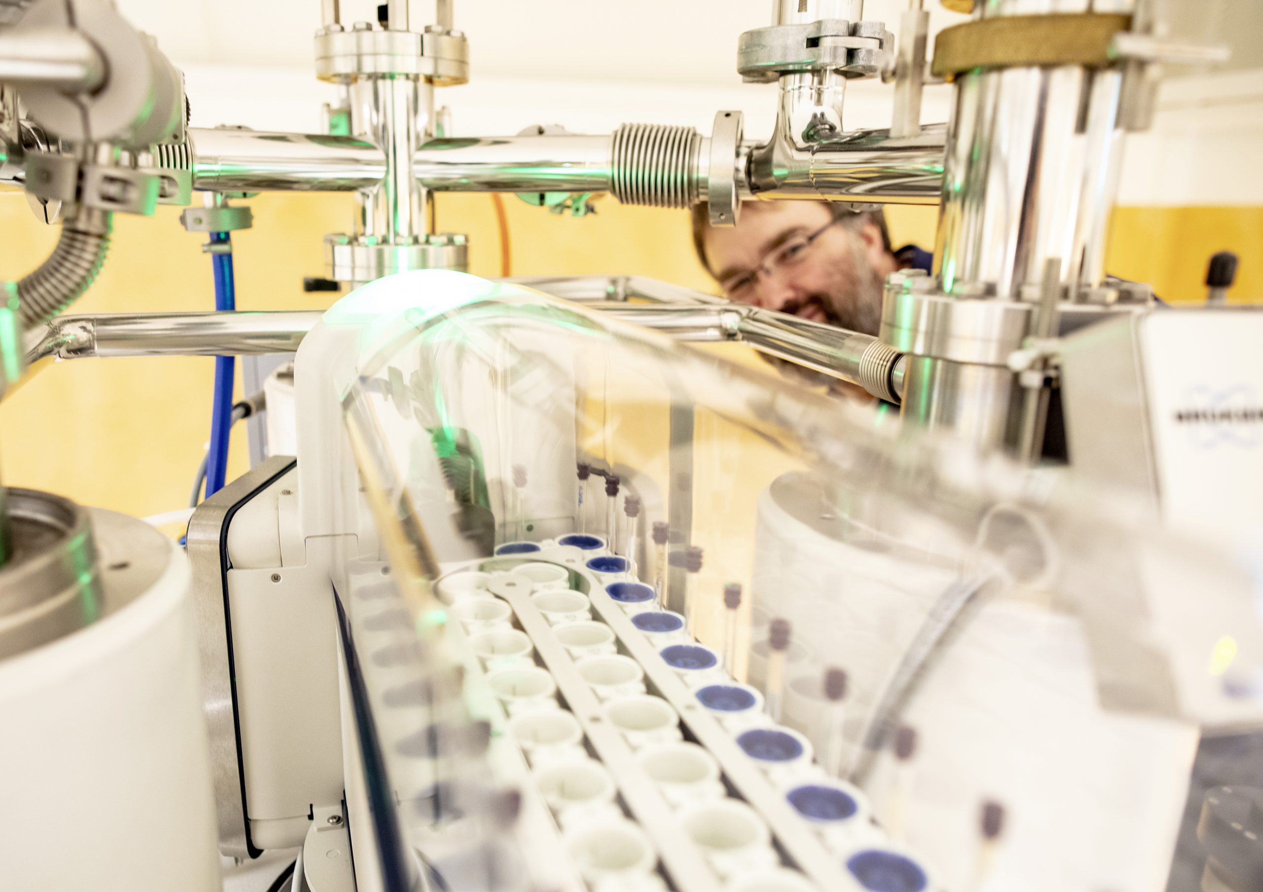
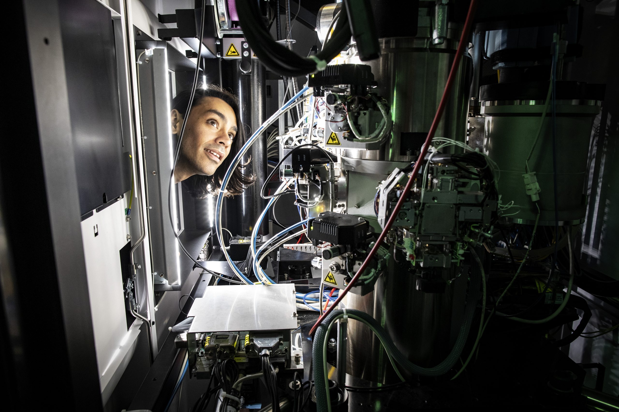
Cryogenic electron microscopy (cryo-EM) is a widely used method for determining 3D structures to near-atomic resolution of suspended biomacromolecules flash-frozen in vitrified ice. Structures can also be determined from the diffraction of electrons by macromolecular samples crystallized on a microscopic scale. With microscopes in Stockholm and Umeå, a network of sample preparation support in Gothenburg, Lund, and Uppsala, and dedicated data-processing specialists, the Cryo-EM unit offers structural biology services and support in:
- Sample characterization by e.g. mass photometry
- Cryogenic grid preparation
- Single-particle cryo-EM data collection
- Microcrystal electron diffraction (micro-ED)
- Data processing and modeling support
Explore more
NMR-based Structural Biology
NMR is a spectroscopic method that will provide insight at the atomic level. Signals are representing individual atoms and therefore one can use each atom in the molecule as a reporter, gaining insight into dynamics as well as specific interactions. NMR is also a method that can be used for structure determination, and is a good choice when Cryo-EM or crystallography is not feasible. Studies can be performed in the liquid state or the solid state.
- Protein structure
- Dynamics
- Protein-ligand interaction
- Protein-protein interaction
- Screening libraries for potiential binders
Explore more
MS-based Structural Biology
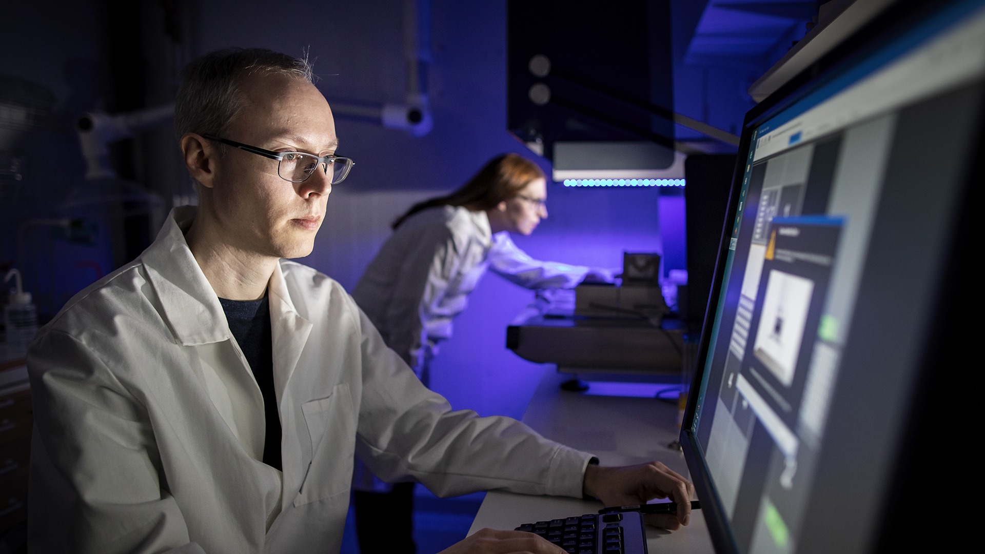
The Structural Proteomics infrastructure unit provides access to cutting-edge equipment and expertise, for analysis of protein interactions and conformational dynamics with mass spectrometry. The unit provides services for all aspects of structural proteomics, including project planning, sample preparation, data collection and data analysis.
- Bottom-up HDX-MS to provide detailed information on protein dynamics and conformation, e.g. identification of disordered protein regions, protein-ligand interaction sites or epitope mapping.
- Cross linking (XL-MS) experiments with a variety of cross linkers to determine topological arrangements in protein complexes and pinpoint where the protein domains interface.
- Denovo Sequencing Provides sequence verification, and allows for characterization of post-translational modifications to ensure antibody integrity and effectiveness.
- Affinity purification mass spectrometry (AP-MS) for analysis of protein-protein interactions (PPIs), or to find interaction partners for a recombinant protein
Explore more
Other Infrastructure Resources
All technologies of protein structural biology require the input of protein samples with high quality, purity and homogeneity. Sometimes specific labelling is necessary, and certain target proteins need to be expressed in a specific host. PPS is a national research infrastructure focused on providing high-quality services in protein production and purification. PPS utilizes a variety of expression systems to enable the production of many different types of proteins and offers expert support for all steps involved in the production process.
- E.coli
- Yeast cells
- Insect cells
- Mammalian cells
- Plant cells
- Cell-free appraoch
ProLinC has been established as a one-stop resource integrating a vast array of (complementary) biophysical techniques and instrumentation; with on-site expertise facilitating experimental design and the interpretation of results…
Critical assessment of proteins and their complexes – stability, purity, binding affinities – enabling optimisation of sample conditions ahead of embarking on costly activities at large-scale infrastructures (cryo-EM, ESS, MAX IV, or Swedish NMR…)
- Access to a diverse range of instrumentation (with technical support and advice if required)
- Consultation with experts: problem solving, experimental design and optimization
- Adept in handling unstable protein systems: IDPs, fibrillation…
Diffraction and scattering techniques employed at synchrotrons yield valuable insights into the structure and shape of macromolecules. Protein crystallography, in particular, enables the generation of high-resolution structural models for proteins and other large biomolecules and their complexes. Small-angle X-ray scattering (SAXS) provides valuable information on the size and shape of macromolecules in solution, facilitating the study of dynamic processes such as complex formation, folding, and fibrillation.
Highly advanced protein crystallography beamlines such as MicroMAX make it possible to study protein processes such as catalysis at a micro to milli second resolution, while BioMAX is ideal for highly automated sample handling and data analysis.
X-ray absorption spectroscopy (XAS) serves as an elegant probe for element speciation in both biological and environmental samples. Notably, it offers detailed insights into the metal centers present in proteins.
- Protein crystallography beamlines BioMAX and MicroMAX
- Small Angle X-ray Scattering beamline CoSAXS
- Hard X-ray spectroscopy beamline BALDER
PReSTO is a software stack for integrated structural biology adapted to high performance compute resources at the National Academic Infrastructure for Supercomputing in Sweden (NAISS) and the local MAX IV compute cluster. PReSTO is working closely with the SciLifeLab Bioinformatics and Integrated Structural Biology platforms to provide efficient and easy-to-use access to supercomputing for structural biology research projects.
Structure Prediction: Advanced protein structure prediction support: LU-Fold is a Lund University-based facility working closely with the SciLifeLab Bioinformatics platform (NBIS), offering national services to help researchers predict protein structures using methods such as AlphaFold2. LU-Fold specialises in high-throughput applications to predict novel protein-protein interactions.
The services include:
- Predicting pairwise binding interactions of a protein of interest with all other proteins in a proteome
- Protein structure predictions of all proteins in a proteome
- Predicting higher order structures of larger complexes
- Predicting the effect of mutations and truncations on interactions
- Workshops and training events
Imaging techniques employed at synchrotrons offer the capability to examine smaller organisms, tissues, and cells across various hierarchical levels, facilitating the study of morphology and organization from the nano to mm scale. By integrating imaging with spectroscopy, researchers can leverage naturally occurring elements without the need for staining. Additionally, tomography provides access to three-dimensional data without the necessity for sectioning. These methods are highly complementary to other forms of microscopy.
- Scanning transmission X-ray microscopy (STXM) beamline SoftiMAX
- Micro tomography beamlines DanMAX and ForMAX
- Nano tomography beamline NanoMAX
- X-ray fluorescence mapping (XRF) nanoscan at NanoMAX
- Infrared (IR) platform with Infrared Nanospectroscopy (AFM-IR) + Optical Photothermal IR (OPT-IR)
- Coming in 2025: Cryo-EMaxiv
The European Spallation Source ESS ERIC is a European project to build the world’s most powerful neutron source in Lund and is expected to open for first users in 2027. Neutron scattering can be used to investigate structure and dynamics in multiple time and length scales, ranging from the atomic to full organisms. Neutron scattering can provide unique information on e.g. hydoigen positions, composition of macromolecular complexes or membrane structure. Dedicated instruments will be available for the different neutron scattering techniques relevant for biology experiments, as well as supporting laboratory facilities for sample preparation and conditioning. Data processing and analysis support will be provided by the ESS Data Management and Software Centre in Copenhagen.
InfraLife is a national project funded by the Swedish Research Council (VR) to maximise the benefits of Sweden’s large scale research infrastructures; MAX IV, ESS and SciLifeLab in partnership with industrial organisations; SwedenBIO and LiF. The aim is to increase knowledge and facilitate access to these LSRI to Life Science researchers in academia, healthcare and industry, while stimulating cross-sectorial collaborations and advancing science.
- Information about the capabilities at MAX IV, ESS and SciLifeLab.
- Stories about Industry cases, events attended and site visits.
- Presentations from Exchange dialogues on Imaging and Fragment Screening.
- Recorded webinars on using LSRI in Drug Discovery & Development.
- Contact details for InfraLife Project Coordinators.