New technique suggest that we only know a fraction about our cellular machinery
Co-led by SciLifeLab researcher Emma Lundberg (KTH), an international team of researchers, have discovered new cell components by using a new technique called Multi-Scale Integrated Cell (MuSIC). The new technique is based on a combination of microscopy, biophysical association and deep learning AI algorithms, and the findings may provide new clues into human development and disease.
Except for viral or bacterial infections, most human diseases are linked to defects in different parts of our cells. Some defects we are born with, such as metabolic disorders from defect mitochondria or genetic mutations leading to disease phenotypes, while others appear during our lifetimes, such as cancer or diabetes type 2.
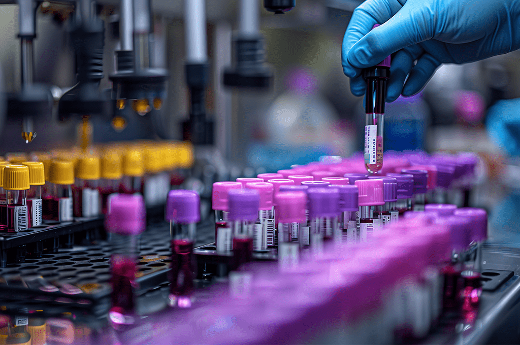
In order to find out what parts of the cell are involved in different diseases, scientists first need to have a list of parts. To study organelles and proteins inside a cell, two techniques are commonly used, microscope imaging or biophysical association.
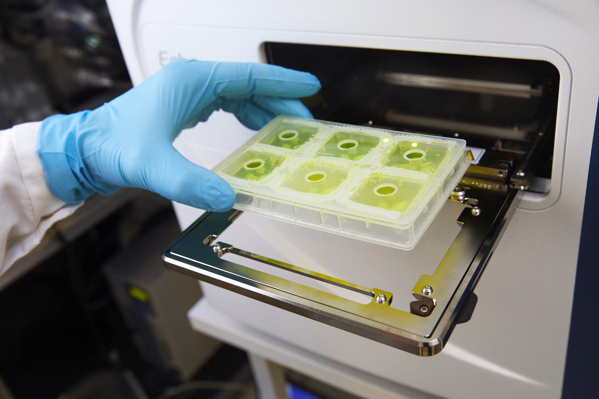
Using microscopy, researchers can visualize some of the smaller organelles, such as mitochondria, by using fluorescently labeled antibodies. In order to study even smaller structures, such as individual proteins or protein complexes, biophysical association is often used. Here, scientists can use antibodies to extract proteins in order to find out what they are bound to.
The problem, however, is that there is a big size gap between the two techniques, with microscopy being able to measure protein distribution at scales from 200 nanometer to 20 micrometer, while biophysical association measures direct interactions from 10 nanometer and downwards.
“How does one bridge this gap from microns to nanometers? That has long been a big hurdle in biological sciences”, says Emma Lundberg, who is also a Director of the Cell Atlas of the Human Protein Atlas.
Now, researchers from Sweden, USA, Belgium and China, led by SciLifeLab researcher Emma Lundberg (KTH) and Trey Ideker from UCSD, have found a solution to the problem. By integrating data from different sources using artificial intelligence (AI) they could assemble a model of functional components in a cell, thus taking a significant leap towards the understanding of human cells. The technique, known as Multi-Scale Integrated Cell (MuSIC), is described in Nature.
“If you imagine a cell, you probably picture the colorful diagram in your cell biology textbook, with mitochondria, endoplasmic reticulum and nucleus. But is that the whole story? Definitely not! Scientists have long realized there’s more that we don’t know than we know, but now we finally have a way to look deeper”, says Trey Ideker, in a press release from UCSD.
The images used in the study came from the Human Protein Atlas (HPA), that started out as a SciLifeLab Research community program (RCP).
“It’s very cool to see how we can use the collection of images in HPA to build a model of a cell and reach novel insights on the function of its underlying system”, says co-author Casper Winsnes, PhD student in Emma Lundbergs group.
In this study, MuSIC revealed approximately 70 components and a least one unknown protein structure, within a human kidney cell line, half of which had never been seen before. The unknown structure turned out to be a new protein complex that binds RNA and is probably involved in splicing.
So far, MuSIC has only been used on 661 proteins and one cell type, which leaves a lot of room for new discoveries.
“The clear next step is to model the proteome of the entire human cell and then move to different cell types, people and species. Eventually we might be able to better understand the molecular basis of many diseases by comparing what’s different between healthy and diseased cells”, Lundberg says.
Artist’s conceptual rendering: UC San Diego Health Sciences
Photo of Emma Lundberg: Daniel Roos