New study reveals protein-level maps of human lung development during early gestation
The lungs are essential for breathing, yet how they develop in early human life has long been elusive. Although recent research has helped us learn more about lung formation, a lot of what we so far know comes mostly from studying animals and is based on transcriptomic data. This has left gaps in our understanding of how lungs develop at the protein level in humans. A new study led by SciLifeLab researcher Burcu Ayoglu, part of the Human Developmental Cell Atlas (HDCA) project, takes a big step forward in filling that gap. It looks at how different types of cells in the human lung grow and organize during the first trimester of pregnancy, specifically between 6 and 13 weeks after conception.
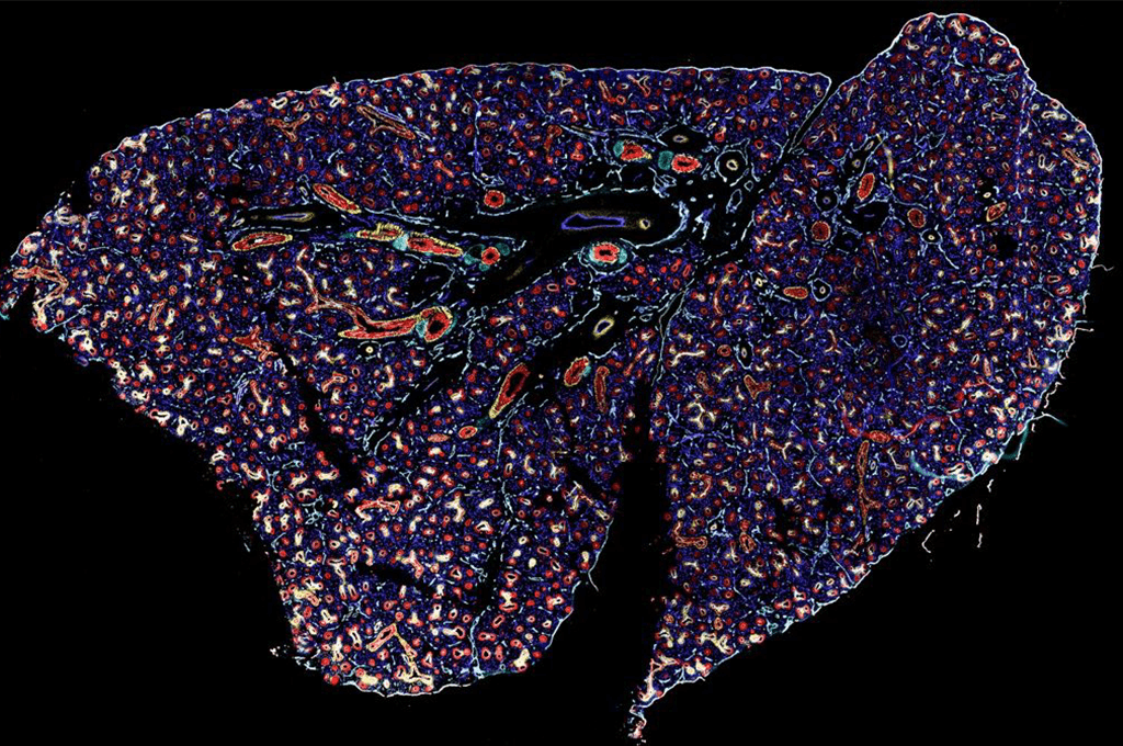
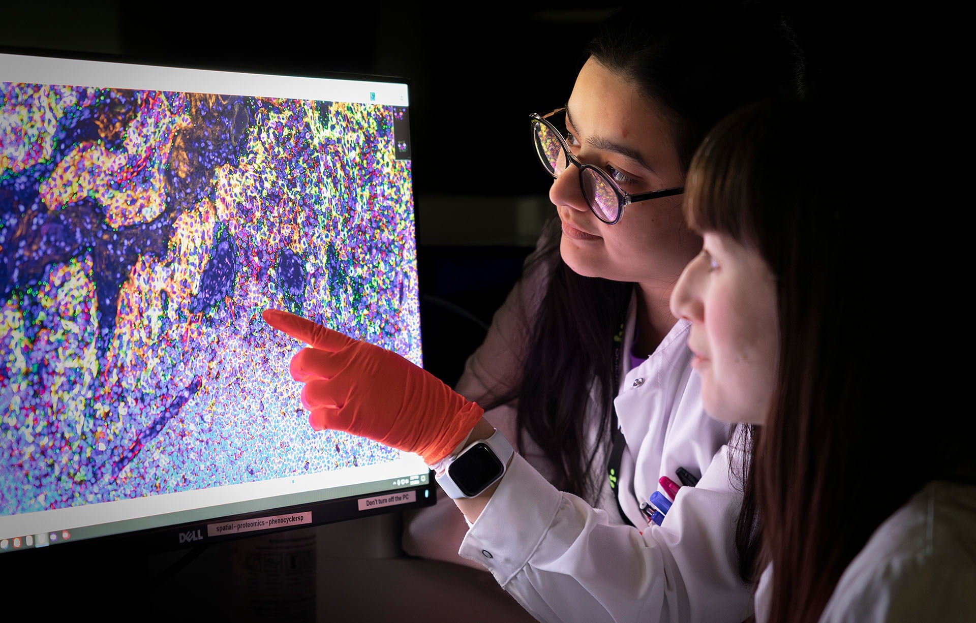
To do this, the researchers used an advanced imaging method with a special set of 30 antibodies that could highlight different proteins. This let them take a close look at over 2 million cells and map them out in detail at different stages of early lung development. This approach is the first of its kind to create such a detailed view of how lung cells are arranged and how they interact as the lung forms, giving scientists a new tool to better understand this process.
One of the key findings was about the growth patterns of different cell types at specific locations in developing lung. The researchers found that cells marked with SOX9, which are located at the tips of the lung’s branching structures, were particularly active. This points to their major role in helping the lung expand.
“By enhancing our understanding of key developmental processes, such as branching morphogenesis and the role of immune cells in angiogenesis, this study provides an important contribution for informing treatments for congenital lung diseases and advancing care for preterm infants,” says SciLifeLab and KTH researcher Burcu Ayoglu.
The imaging-focused approach also gave the team a possibility to identify distinctive spatial arrangements among cell types within the tissue. Notably, they found that a type of immune cells called macrophages often gathered around developing blood vessels in a ring-like pattern. This pattern had been seen in animal models before but hadn’t been well-documented in humans. This finding hints that macrophages might help shape blood vessels as they grow, suggesting they do more than just support the immune system during human lung development.
Burcu Ayoglu also talked about the support from SciLifeLab and how it impacted the project: “SciLifeLab’s proximity to gynecology clinics in the Stockholm region, along with its links with experts who have established robust, ethical protocols for routine sampling of embryonic tissues, has been essential to this project. The SciLifeLab Data Centre has also provided vital support by hosting a publicly accessible portal that allows researchers worldwide to explore datasets from all single-cell and spatial modalities on the same sample cohort, further enhancing the project’s reach and impact.”
In the end, this study provides an important, openly available resource: a single-cell, protein-level map of how the human lung develops in the first trimester. This detailed map can be explored online and could help scientists learn more about how early lung development affects health later on, including how certain congenital lung conditions might begin.
DOI: https://doi.org/10.1038/s41467-024-53752-x
What is the Human Cell Atlas (HCA) initiative?
The Human Cell Atlas is an international research consortium focused on creating a detailed map of all cell types in human body. By constructing a comprehensive reference map of human cells, HCA aims to transform our understanding of health and disease, providing insights that could drive major advancements in healthcare and medicine globally.
Human Developmental Cell Atlas (HDCA)
HDCA (hdca-sweden.scilifelab.se) is a Swedish initiative within the larger HCA project, aiming to build a comprehensive molecular map of human prenatal development, with a primary focus on the first trimester when the most critical stages of embryogenesi occur. HDCA uses state-of-the-art technologies established at SciLifeLab, including single-cell RNA, protein and spatial techniques. The project is organized, located and managed at SciLifeLab.


