Industry case: Early 3D kidney diagnostics development with Magnephy using SciLifeLab advanced light microscopy
The biotech company Magnephy is developing novel methods to analyze tissue biopsies using optical 3D microscopy and sample clearing, a workflow with potential for whole tissue diagnostics and in-situ drug screening. The company has its roots in research at KTH, KI and SciLifeLab, and today, Magnephy use SciLifeLab’s infrastructure for advanced light microscopy support.
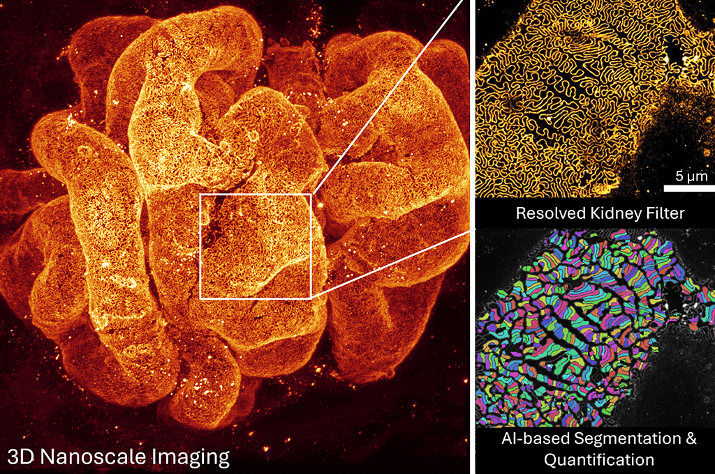
Magnephy apply state-of-the art optical 3D microscopy and AI-supported image analysis to accurately quantify pathologies in whole biopsies and to support pharmaceutical drug development.
SciLifeLab has, since 2014, been part of Magnephy’s development journey until the company spun off from KTH Innovation in 2023. Previous optical microscopy and sample preparation research within SciLifeLab, KTH and KI, laid the foundation for the 3D methodology, which is now also being validated to better diagnose kidney diseases clinically. Chronical kidney failure is often discovered late with reduced success in interruptive treatment. Magnephy’s technology can enable earlier automatic quantitative 3D detection of pathological changes and can, therefore, play a crucial role in renal precision medicine support.
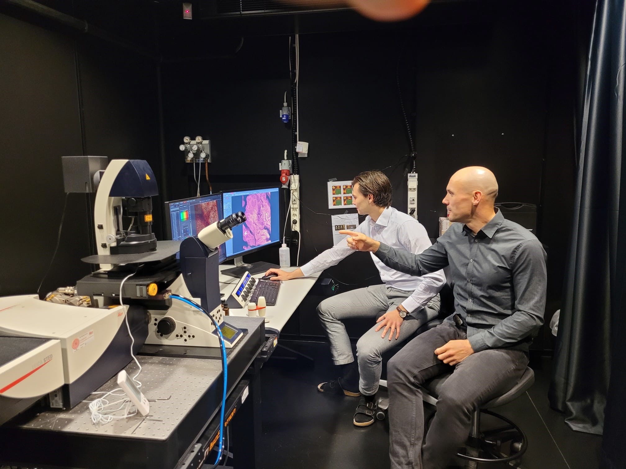
“That a spin-off company can rent time on our nationally unique advanced light microscopy equipment and get access to SciLifeLab expertise has been extremely valuable during their start-up phase,” says Hans Blom, KTH, and Head of SciLifeLab’s unit for integrated microscopy technologies in Stockholm.
Magnephy collaborates with both Swedish and international pharmaceutical companies to analyze tissue biopsies exposed to drugs. One can use their 3D optical method to study how drug localization correlates to disease curing automatically with AI-assisted image analysis.
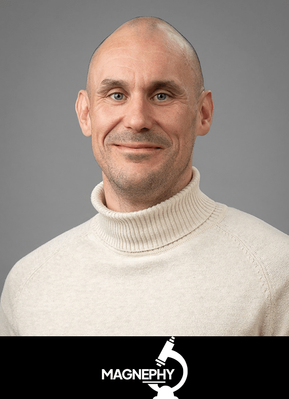
“Our data-rich method allows us to detect pathological changes at a very early stage using whole 3D biopsy imaging. Hence providing the opportunity to interfere and possibly reverse or stop disease progression. We can not only identify early changes better than current 2D protocols but also automatically assess whether diseased states are reversed by a given treatment on the nanoscale,” says David Unnersjö, CTO of Magnephy.
With service support from SciLifeLab’s infrastructure in advanced light microscopy, Magnephy continues to drive development forward. The next step is to deepen collaborations within healthcare and further improve the precision diagnosis and treatment of kidney diseases using optical 3D microscopy. The company is already working with pathologists and clinicians to validate the method for hospital workflows. “We need to compare and validate our method with current ‘golden standards’ used clinically in kidney pathology. Our whole biopsy 3D method could thereafter be integrated into building a more automated procedure for generating very useful diagnostic support,” says David Unnersjö
“Why kidney samples? Imaging it better optically in 3D has been a focus at KTH and KI since the mid-1990s. Thus, driving advancement in optical microscopy, fast and easy whole kidney biopsy preparation, and automatic AI-assisted image analysis, our current research field, we knew how important improved 3D diagnostics of pathological changes detected early and at the microscale would be. The 3D optical workflow is further not tissue specific but has been tested on for example liver, lung, brain, and intestine biopsies as well,” Hans Blom explains.
For further information and access to SciLifeLab infrastructure, please contact Hans Blom, Head of SciLifeLab’s unit for integrated microscopy technologies in Stockholm.
Want to know more?
Contact:
Magnephy
David Unnersjö
CTO
Magnephy website
david.unnersjo-jess@magnephy.com

SciLifeLab
Hans Blom
Head of Unit: Integrated Microscopy Technologies
IMT Unit Website
hblom@kth.se