Spatial Proteomics Method of the Year: “great acknowledgement”
Nature Methods has chosen spatial proteomics as Method of the Year “for its critical role in revealing the organization of complex tissues”. We asked Associate Professor Charlotte Stadler, Co-Director of the Spatial Biology platform and Head of the Spatial Proteomics unit at SciLifeLab for some insights into the method – or methods to be precise.
What is spatial proteomics?
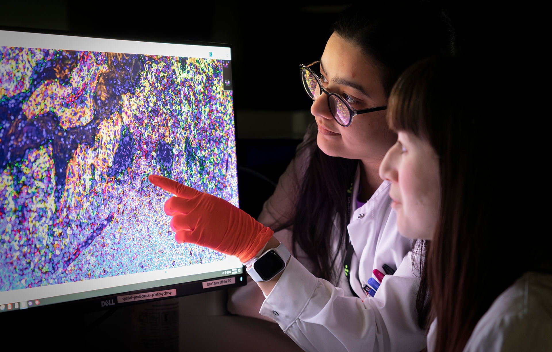
Spatial proteomics is an umbrella term of immunohistochemical methods used to profile complex tissues. While imaging techniques including immunofluorescence have been around for a long time, recent advances now allow for tens – or even a hundred – proteins to be analyzed within a single tissue section. This allows for much deeper information to be obtained.
What’s the strengths of spatial proteomics?
The inherent imaging itself and single cell resolution, and that the tissue structure is retained. As such, cells are defined and studied within their natural context. Therefore, the link between tissue architecture and function can be explored in a spatial context.

Why do you think spatial proteomics has been chosen as method of the year?
Because of the strengths mentioned, but also the recent advancements in the imaging field that now allows for highly multiplexed experiments to be done. Before they were limited to studies of a few markers in the same sample. Now there are several approaches to capture a multitude of different protein markers, and also together with transcripts or other spatial modalities. One should also not forget that the proteins, which is the readout from these assays, are the functional entities of our cells and hence mapping their expression can tell us more about the functional states of the cells.
What’s next for spatial proteomics?
The next steps for spatial proteomics is the continuous integration with other omics types, with equally good resolution and image quality. An interesting new combination that has not been explored so much yet is the combination of proteins and small molecules such as metabolites and lipids.
In the Spatial Proteomics Unit at Scilifelab we are also working to complement the multiplexed imaging of proteins with in situ proximity ligation assay (PLA), to look at protein interactions and cell signalling within the tissue microenvironment.
Also, for the field in general, moving from 2D to 3D will lead to yet more information of tissue and cellular structures. At the same time, much focus is put on bringing these methods closer to clinical practice, they have huge potential to contribute to better diagnosis and treatment selection.
What services does SciLifeLab offer within spatial proteomics?
At SciLifeLab we offer full project support to generate spatial proteomics data using a portfolio of different technologies including Akoya Biosciences, Inc. PhenoCycler Fusion and Lunaphore COMET – Lunaphore is a Bio-Techne brand, and various assays compatible with the new instrument from Thermo Fisher Scientific, Evos S1000. If users are ready to make use of these informative methods but lack the know-how and instruments, we are happy to discuss the project and offer support.
“I am very happy to see this great acknowledgement for immunohistochemistry based methods within Spatial Omics, used for profiling the functional entities of cells – proteins!” concludes Charlotte Stadler.
Lean more at the SciLifeLab Spatial Proteomics unit page and in the article from Nature Methods


